Philodendron Anatomy: Complete Botanical Guide to Plant Structure
What if I told you that every Philodendron leaf presents with mathematical accuracy at precisely 137.5 degrees from its predecessor — the same “golden angle” found in sunflowers and nautilus shells? This botanical masterpiece is more than gorgeous; it’s an evolutionary triumph that has enabled these tropical climbers to reign supreme over rainforest canopies for millions of years. Knowing this complex arrangement explains why Philodendrons have evolved into the most beautiful of indoor gardening implements — and how their complex anatomical systems have evolved to work in some of nature’s toughest environments.
Architecture of the Roots: The Building Block of Climbing Success
Philodendrons also display one of the very advanced root systems of the plant world with both aerial and subterranean features which do very different and yet complementary jobs. This two-sided-root system is millions of years of evolutionary improvement that has facilitated such plants to grow as forest floor dwellers, and later become epiphytes (canopy).

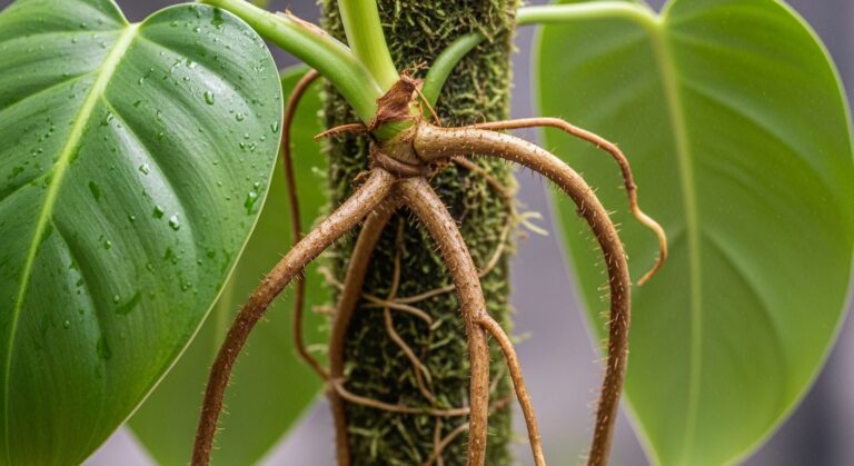
Specialized Function of Fibrous vs Aerial Roots
There is striking dimorphism in the root system, where feeding roots and anchor roots grow on the same plant at the same time. Feeding Roots, 1 to 2 m long in the typical adult plant, are dug into soil layers, capturing water and essential nutrients. These roots are positive hydrotropes growing toward water; they grow at a high accuracy toward water from its sources, and negative heliotropics, showing off of very few in the distance due to strong light exposure are visible.
Anchor roots, on the other hand, are shorter and more common and contain specialized epidermal cells that generate root hairs for better attachment to bark surfaces. These structures are able to perceive chemical gradients in the surrounding environment, enabling them to learn optimal site(s) of attachment to host trees. The transition between root types occurs at different nodes but feeding roots are usually found at every third node in climbing species PMC.
Cellular System and Specialized Equipment
Underneath this epidermal skin, there are some of the characteristic sclerotic hypodermis, cylindrical tubes, one to five cells that add strength to the skin structure. These cells lignify and form protective walls for pathogens while also retaining their plasticity to flourish. The cortex is composed of three parts: outer, medial and inner spaces, with cell organization and function-specific structures.
Resin-connecting ducts run throughout the cortical area and form a network of schizogenous spaces, enclosed by epithelial cells. These structures, which belong to the Araceae family, release protective compounds to help ensure chemical communication between plants. The endodermis, at early stage III development, has an asynchronous maturation path and in this third stage, its Casparian strips are very prominent and it controls the transport of water and nutrients into the vascular system.
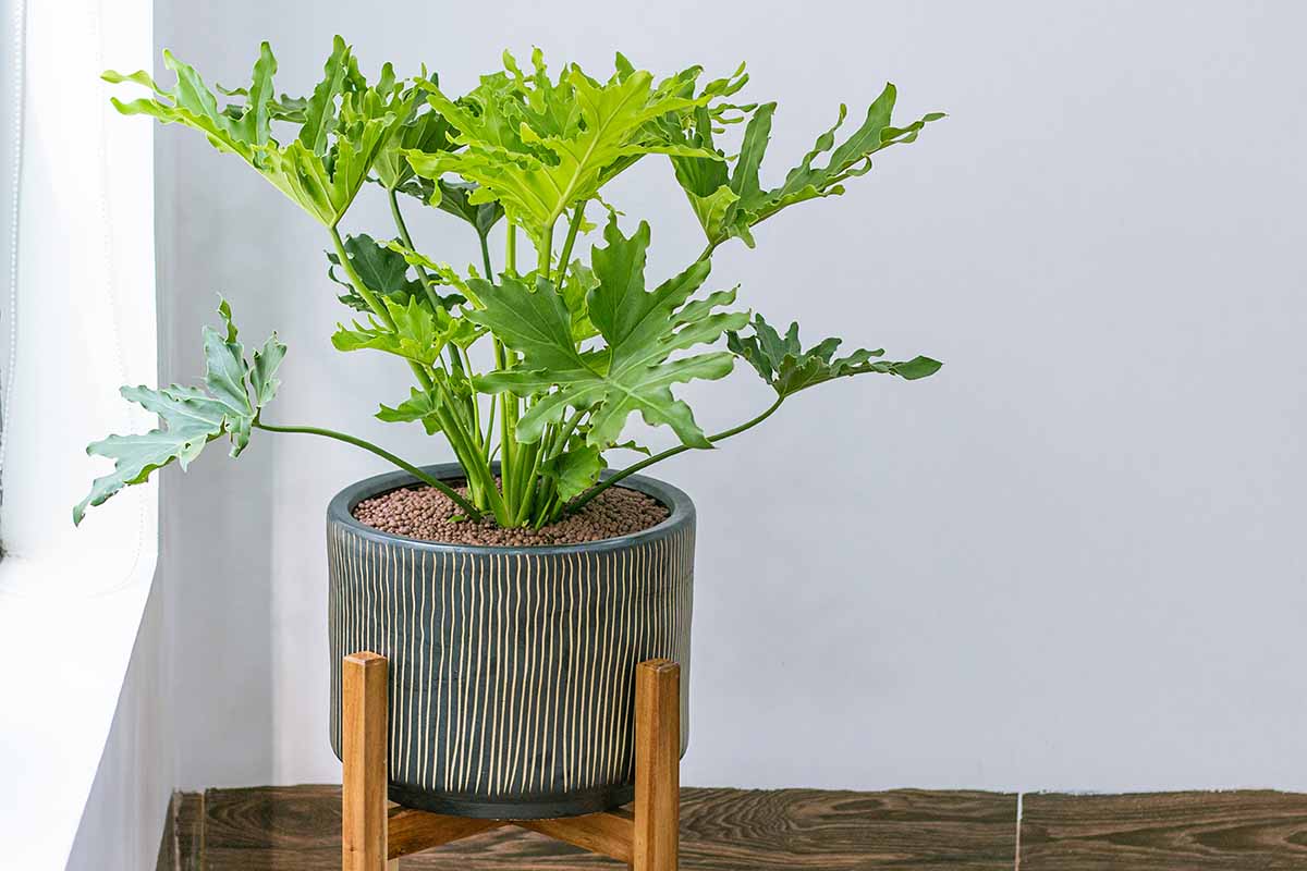
Stem Structure and Growth Pattern: Plant World Engineering Marvels
Philodendron stems are architecturally stunning devices that are both stiff and flexible. Stems differ greatly among the three overall growing habits: climbing, hemiepiphytic and self-heading.
Structural Structures and Mechanical Assistance of Vascular Structure
The vascular system consists of collateral bundles arranged in a complex configuration, with pairs of long and short strands of phloem arranged one after another. Protoxylem elements show spiral forms of thickening, and metaxylem vessels reach 50–80 micrometers in mature stems. Climbing species demonstrate thicker pericycle layer cell walls that are helpful with mechanical support in vertical growth.
In climbing trees the number of stem diameters varies, with a maximum being 5-15 millimeters and in terrestrial varieties exceeding 30 millimeters (eg, Philodendron bipinnatifidum). Growth rings appear in old specimens, where each ring was an early stage for growth under ideal conditions. The pith also has a large internal aerenchyma tissue — specialized cells with big intercellular spacing that facilitate gas exchange and buoyancy in underwater compartments.
Mathematics of Growth Patterns and Circadian Precision
Philodendron growth is guided by mathematical principles that optimize resources and efficiency to reduce complexity. For climbing species, the internodal distance has an average of 2-8 cm, with carefully spaced distances to avoid self-shading while maintaining structural integrity. Even though it grows very closely together (so there’s light in the air), it is still flexible, adapting its structures to weather.
The stems grow determinate in juveniles and grow indeterminate when adult. The change is associated with hormone shifts, mainly increased auxin synthesis at apical meristem. Influenced developmental movement between patterns of growth can be very sudden, possibly during a single growing season even by the time selected under ideal environmental conditions.
Variation in Leaf Morphologies: The Wonders in Plants is Nature’s Most Famous Game
Philodendrons’ leaf morphology is a classic example of developmental plasticity at the zenith of plasticity in the plant kingdom. The transference of juvenile to adult leaf morphology (a heteroblast form) involves profound alterations in leaf structure, venation patterns and functional morphology.

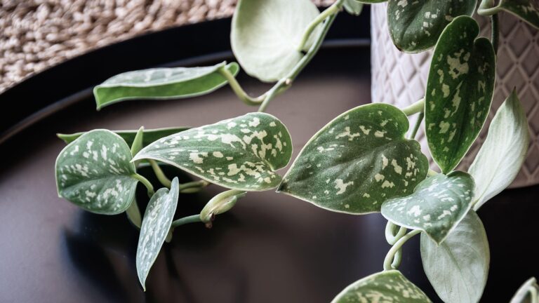
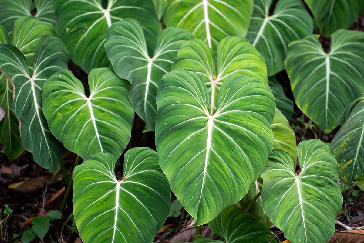
Heart-shaped Start-up of Juvenile Leaves
Juvenile Philodendron leaves appear as basic heart-shaped leaves with very small lobes and entire margins. These leaves tend to be 5-15 cm in length and have cordate bases with acute to acuminate apex. The lamina thickness ranges from 165-400 micrometers, in addition to a bifacial structure adapted for maximizing light harvesting in the understory spaces.
Epidermal cells have polygonal shape with straight anticlinal walls, and cuticle layer is measuring 2.5-5.3 micrometers thick on adaxial plane. On the abaxial surface, stomatal density is on average 4.66-21.53 per square millimeter and is significantly reduced on the adaxial surface in order to reduce wastage in shaded conditions. Mesophyll structure is of 1-3 layers of palisade parenchyma with spongy cells with intercellular areas for gas exchange.

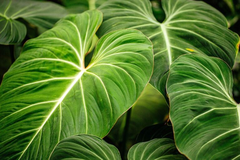
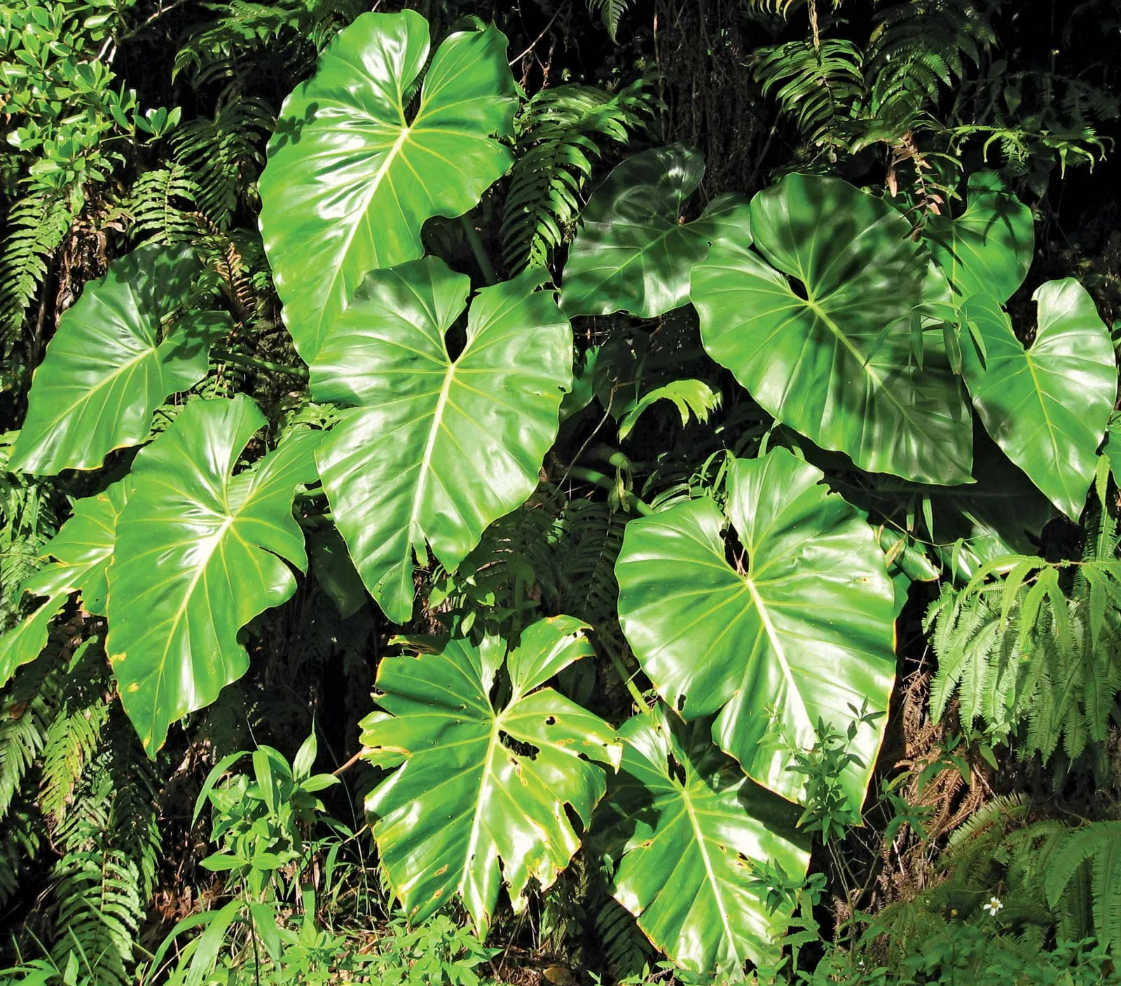
Striking the Right Sides: Canopy Life Mechanics of a Split and Fenestrated Mature Form
Leaf Form morphs undergo dramatic changes when plants mature and climb to brighter conditions. Deeply lobed fenestrated leaves appear, with splits extending up to 75% of the length of the lamina section. These changes have different roles — dampening wind resistance in exposed canopy positions, prohibiting the build-up of water, which could promote the growth of fungi, and maximizing light interception by minimizing the leaf area of interest.
Fenestration is initiated by the generation of specific cells at specific locations, through gene-coded programs that depend on physiological signals from hormones. These cells die as a result of programmed cell death and form holes or splits that track mathematical patterns. The 137.5° golden angle is responsible not only for the arrangement of leaves but also for fenestration positions, which results in light distribution throughout the canopy.
Cellular Structure and Adaptations During Ageing
At rest, hypodermal cells form in 1-4 cell layers beneath the epidermis that protect the cells against UV radiation and mechanical damage. Collenchyma cells develop continuous bands with angular thickening, providing mechanical support for higher leaf sizes, up to as much as 1 meter in diameter.
The vascular system becomes complex with the maturity stage including collateral bundles with fiber caps supporting both xylem and phloem tissues. Air cavities arise within the midrib and are cleaved by uniseriate ground tissue in the midrib with uniseriate areas holding structural integrity yet minimising weight. The modifications can enable mature leaves to reach up to 3 feet across by protecting their structural integrity in windy canopy situations.
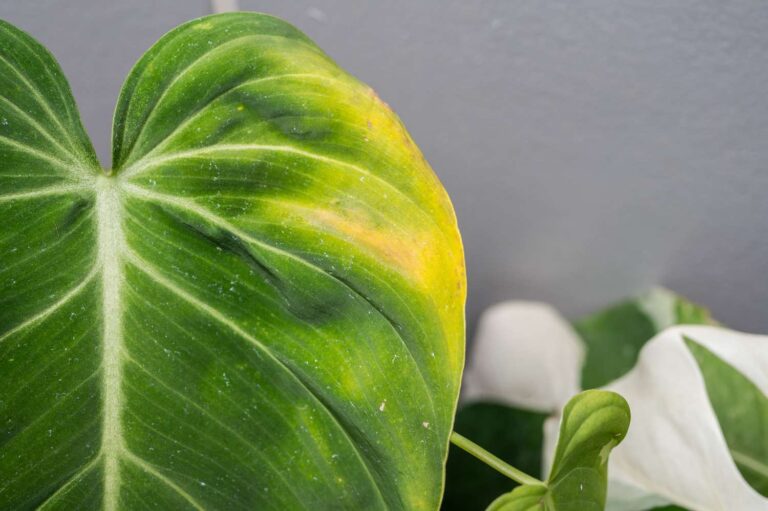
Venation Pattern and Cellular Structure: The Transportation Network
Venation patterns in Philodendron leaves indicate complex transportation systems that progress over the course of plant growth. These are not primarily functional but are useful for species identification and phylogenetic analysis as they can also be diagnostic.

Venation Architecture – Primary and Secondary
The primary venation is usually pinnate, with the secondary veins branching from the midrib, approximately at 45-75 degrees. These veins are separated by fiber reinforcement to prevent collapse under negative pressure during transpiration and are 150–500 micrometers in diameter. The midrib includes multiple vascular bundles present structurally and protoxylem and metaxylem vessels are noted to feature spiral and scalariform thickening.
Secondary vein density increases significantly from juvenile to mature leaves; spacing can be reduced from 2-3 millimeters to less than 1 millimeter between adjacent veins. This higher density is related to high transpiration rates and increased photosynthetic capacities in mature canopy leaves. There are particular tracheary components found at the vein endings; these enable water transport into mesophyll tissues.
Tertiary and Quaternary Networks
The formation of higher-order venation generates complicated networks that provide an optimal contact area with mesophyll cells on the surface area. Secondary veins have areoles, where the areoles are generally between 0.1-0.5 square millimeters in area, and tertiary veins can connect secondary veins at angles typically of 60-90 degrees.
This density network differs markedly among species that are adapted to vary in light; shade-tolerant species have a lower density when compared with sun-tolerant species. The quaternary veins feature free-ending veinlets that run as deep as mesophyll and provide effective watering throughout the lamina. These terminations invariably have well-defined tracheary elements with lower wall thickness to allow for rapid ingress of water into adjacent cells. There is hydraulic connectivity throughout the whole network via pit membranes for selective transport of water and for preventing air bubble propagation.
Stomatal Distribution and Performance
The distribution of stomata conforms to exacting patterns which allow for easy gas exchange and at the same time avoids wasted water. Young leaves exhibit stomata, which are found mainly along the abaxial midrib and margins with densities of 20-25 per square millimeter in ideal conditions.
In mature leaves, they are more evenly distributed and have stomatal densities between 8-15 per square millimeter depending on the site of origin and environment. Morphology of guard cells vary between species: 21-34 micrometers in length and 8-14 micrometers width. Subsidiary cells exhibit brachyparacytic, brachypara-tetracytic, and brachyparahexacytic arrangements. These arrangements modulate stomatal function and response to environmental stimuli, and a few species show rapid stomatal closure upon drought stress.
Flower Development and Inflorescence Structure: The Reproductive Marvel
Philodendron inflorescences are one of nature’s most complex reproductive structures, combining mechanical precision with biochemical intricacy. The inflorescence develops through a sequence of specific stages which has its own evolutionary significance in reproduction.
Spadix and Zonal Organization
The spadix is the central reproductive axis containing flowers organized into several zones that inhibit self-pollination and can maximize reproductive performance. Female flowers occupy the basal 30% of the spadix length, and have 7-10 carpels per flower with horseshoe-shaped primordia that arch inward. These are protogynous flowers (stigmas) receptive 24 to 48 hours before stamens release pollen.
Male sterile flowers constitute an intermediate zone between 10 and 15% of the spadix length, establishing a biochemical boundary between the male and female regions. These flowers produce rich lipids as food rewards for pollinating beetles and scents that attract specific pollinator species; thus feeding on them. In the sterile zone also resin compounds, unique to Philodendron, are produced, making the pollinators sticky, aiding pollen transfer between the plants.
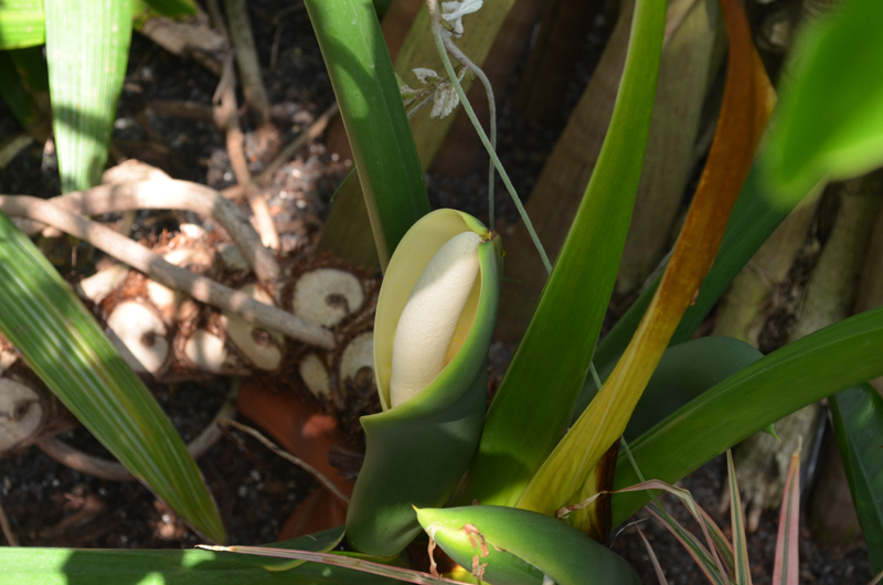
Development and Thermogenic Function of the Spathe
The spathe grows as a modified leaf that encases and protects a developing spadix. The spathe opens up during anthesis, exposing the spadix but at a 45-degree angle that maximizes thermogenic heat dissipation and pheromone dispersal. This position of cells aligns precisely based on their growth profile, especially the expansion in the pulvinus region, where cells expand unevenly to orient them correctly.
Thermogenesis is a remarkable adaptation, with spadix temperatures rising 15-20°C higher during peak reproductive activity compared with ambient conditions. This heat is generated by alternative respiratory pathways in mitochondrial cells, consuming stored lipids at rates equivalent to a hummingbird’s metabolism. The rise in temperature favours volatilization of appealing compounds and also provides thermal comfort for beetles bearing pollinators.

Pollinator Biology and Timing of Reproduction
Controlled pollination depends on intricate timing processes to ensure the success of cross-pollination. Female anthesis occurs entirely on the first night of inflorescence opening and is maintained for 12-18 hours until stigmas remain receptive. Male anthesis is 24-48 hours later, corresponding with maximum pollinator activity levels. This timing avoids self-pollination while optimizing pollen transfer between individuals.
There’s a unique evolution pattern in Philodendron that is resin production. As a result, they coat pollinating beetles so they stick and ensure that pollen adheres on trees while they travel between inflorescences. Additionally, the resin embodies antimicrobial features, which protect maturing seeds from pathogen takeover leading to reproductive success in the competitive rainforest.
Fruit Creation and Seed Generation: The Future Science of Fruit Growth
The formation and development of the philodendron fruit also requires quite complicated biochemical features maintaining seeds that they can successfully disperse away. The shift from flower to fruit needs precise hormonal signaling that orchestrates cellular differentiation and resource allocation.
Development and Maturation of Berry
After the ovary becomes a berry due to successful pollination, it is made up of several small seeds. Berry development takes 2 to 12 months, depending on species and in different environmental conditions, and most of the tropical species require 4 to 6 months for complete maturation.
As it grows, the spathe becomes larger for the growth of berries, but this will shield them from harmful elements and herbivores while shielding from stress and environmental change. Berry colouration is obtained by pigments in a complex process and white and green appearance of white to green are not only the feature of the fruit but also appears in many species, orange or orange-yellow are used and greenish color of some plant species that mature. This color shift is associated with higher sugar contents and volatile compounds production by seed dispersing animals as the fruit begins to grow. The mesocarp also contains calcium oxalate crystals that will protect it from being eaten too soon as the fruit develops.
Structure of the Seed and Germination Requirements
Philodendron seeds are adapted well to rainforest ecosystems. Seeds are 1-3 mm in diameter and are endowed with endosperm tissue to feed the developing seedlings in their early stages. The seed coat contains structures that enable the seeds to adhere to animal dispersers and protect them from mechanical damage.
Germination requirements vary widely among species: in some 75-85°F ranges for the seedling but in others, temperatures as low as 65°F may be sufficient. Wetting conditions need consistently high (at 80-90% relative humidity) in order for germination. Many species have delayed germination capabilities, so seeds can survive up to several months until conditions suitable for germination develop.
Dispersal Pathways and Environmental Significance
Seed dispersal relies on several mechanisms that help sustain genetic diversity and facilitate colonization. The primary route of dispersal is through consumption by animals, and bats, monkeys and birds are primary vectors. These species feed on berries and deposit seeds at distances (e.g. 100 meters to several kilometers) from the parent plants, which encourages gene transfer between populations.
Seed dispersal is mediated by ants to the tiny-seeded species. Ants selectively seek out structures found on seed surfaces containing lipid-rich substrates. That is, this myrmecochory dispersal method ensures that seeds arrive in nutrient-rich microsites, increasing the chance of successful seedling life. This blend of dispersal methods helps drive the unique success of Philodendron in a range of tropical ecosystems.
Conclusion
Philodendron plants possess anatomical complexity spanning millions of years of evolutionary refinement, resulting in intricate forms of functional excellence. The mathematical precision of the arrangement of the leaves at the golden angle or the complex reproductive strategies that are used to achieve thermogenesis or chemical transfer from plant to insect indicate engineering principles that have inspired botanical research and horticultural applications.
The anatomy of Philodendron plants reveals everything about them, including how plants adjust to life as it happens (in a process many organisms know through biology and biology themselves). Such a delicate interplay of structural innovation and functional efficiency, so crucial to successfully functioning in the wide range of tropical environments, leaves them ideally suited to study in science and cultivation.
Key Sources:
Anatomy of the adventitious roots of Philodendron (Araceae) | PMC
Anatomy of the adventitious roots of Philodendron | ResearchGate
Philodendron Plant Guide | NC State Extension
Philodendron Pollination and Thermogenesis | In Defense of Plants